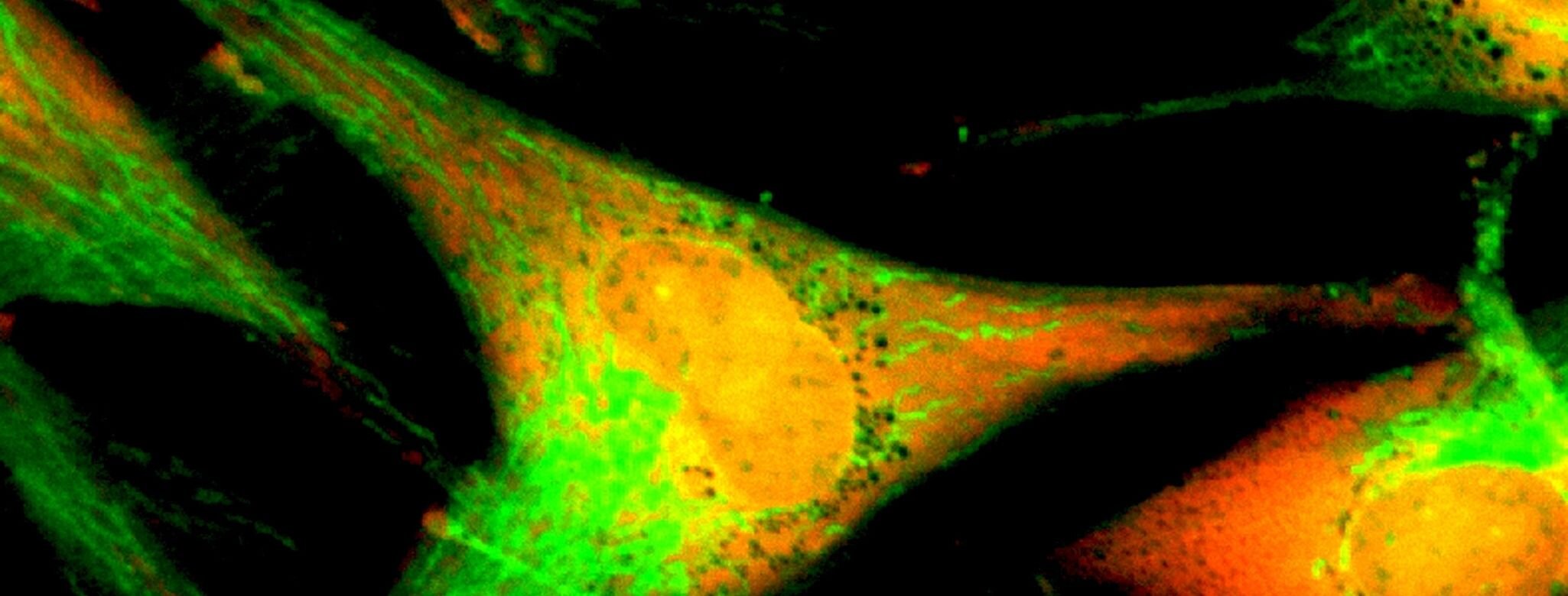
Skin - Melanocytes
Melanocytes are found in the basal layer of the epidermis as well as in the hair follicles and produce a pigment called melanin.
a. Epidermal Melanocytes
The ratio of melanocytes to keratinocytes in the epidermal basal layer is 1: 10 , with around 1200 melanocytes found per mm2 of skin. When melanocytes are seen under a microscope, it is observed that each cell contains organelles called melanosomes that produce two types of melanin pigment compounds : pheomelanin and eumelanin. The ratio in which these pigments are present in the skin, result in the diversity of human skin colour. In addition to providing the skin with its colour, melanin helps in skin protection when exposed to the harmful UV rays from the sun. The most common evidence of this is the development of a tan, when exposed to sunlight for extended periods of time, where melanin production is increased transiently, in response. If melanocytes aggregate together to form a patch, it may result in visible birthmarks, freckles or age spots on the skin.
Compared to pheomelanin, eumelanin is more effective in protecting the skin against the harmful effects of UV radiation and free radicals. This is why there is a 30-40-fold higher risk of skin cancer in people who have lighter skin with reduced eumelanin, when compared to people with darker skin.
If melanocytes undergo a malignant transformation, that is, if they undergo a change from normal benign cells to atypical, neoplastic cells, with hyperplasia and hypertrophy, they form melanomas, which is a type of skin cancer. This may occur on the surface of the skin, in the irises of the eyes and very rarely inside the nose or throat. The transformation is largely attributed to prolonged exposure to UV radiation and can be treated successfully if caught early. However, evidence shows that it is an aggressive cancer that tends to spread beyond its primary site. The incidence of melanomas has risen by 4-6% annually. While it still represents less than 5% of all cutaneous malignancies, melanoma accounts for the majority of deaths due to skin cancer.
b. Hair Follicle melanocytes
Melanocytes are located in the proximal bulb of each hair follicle as well as near the hair shaft, in the sebaceous gland. The ratio of melanocytes to keratinocytes in these areas is 1:5. A three part hair melanin unit or follicular melanin unit, made up of fibroblasts, keratinocytes and melanocytes is formed. Structural and functional interactions between these cells, allows melanin to be transferred from melanin granules into keratinocytes, forming coloured hair shafts. The amount and colour of the melanin is determined genetically for individual people. This is due to the balance maintained between the brown-black eumelanin and the yellow-red pheomelanin.
Human melanocytes are distributed not only in the epidermis and in hair follicles but also in mucosa, cochlea (ear), iris (eye), and mesencephalon (brain) among other tissues.
Disorders affecting Pigmentation
Unequal distribution of melanin may cause pigmentary disorders like :
Hyperpigmentation : These are areas where excessive melanin is present and are seen as dark areas on skin. These can be transient, due to sun exposure, residual acne scars or skin infections like folliculitis. During pregnancy, hormonal changes causes darkened, discoloured patches on the face, known as chloasma or the mask of pregnancy.
Birth control pills, hormone therapy, stress or thyroid disease can give rise to dark patches on the skin, known as melasma.
Hypopigmentation: These are patches of skin that are lighter than the surrounding skin tone. While healed scars from injuries or burns may cause focal areas of hypopigmentation, certain genetic conditions like albinism, are also responsible. Auto-immune conditions and overactive thyroid glands may destroy melanocytes in certain areas of the skin , leading to a patchy loss of pigment, known as vitiligo. Fungal infections like Tinea versicolor may also cause hypopigmented lesions on the skin.
It is interesting to note that melanocytes and their pigment manufacturing melanosomes are not only studied in human beings. Archaeologists conduct tests on remnants of melanocytes preserved in fossils to find clues that could hypothesize the colours of the dinosaurs that lived millions of years ago!
References
Cichorek M, Wachulska M, Stasiewicz A, Tymińska A. Skin melanocytes: biology and development. Postepy Dermatol Alergol. 2013;30(1):30-41. doi:10.5114/pdia.2013.33376
Matthews NH, Li WQ, Qureshi AA, et al. Epidemiology of Melanoma. In: Ward WH, Farma JM, editors. Cutaneous Melanoma: Etiology and Therapy [Internet]. Brisbane (AU): Codon Publications; 2017 Dec 21. Chapter 1. Available from: https://www.ncbi.nlm.nih.gov/books/NBK481862/ doi: 10.15586/codon.cutaneousmelanoma.2017.ch1
Yamaguchi Y, Hearing VJ. Melanocytes and their diseases. Cold Spring Harb Perspect Med. 2014;4(5):a017046. Published 2014 May 1. doi:10.1101/cshperspect.a017046
https://www.healthline.com/health/melasma#symptoms
https://www.nhs.uk/conditions/vitiligo/
https://www.healthline.com/health/skin-disorders/hypopigmentation#causes


